
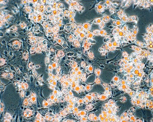
This is an image of adipocytes (fat cells) that have been stained with a compound called Oil Red-O. Oil Red-O binds to lipids (oil) and stains them red. This photo was taken with a light microscope attached to a digital camera. This is from an experiment from when I worked with stem cells. These fat cells were made from other cells that I treated with some steroids, vitamins, and some extra carbs (I'm not kidding) and after a few days...Voila! Fat!
Now some of you might be thinking why are stem cell researchers making fat and where can I donate mine for money? Well in studying the development of fat it is possible to understand obesity and other disorders such as lipodystrophy (a lack of enough fat).
All that aside when I saw these cells after I had "made" them I thought it was actually quite beautiful. It was one of those days where I was glad to be working in science. Mostly because my experiment worked but partly because of the things I get to observe.
Its not art per se but its nice to look at and I made it.
There are even more sophisticated ways of imaging cells.
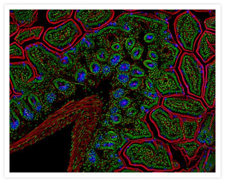
This photo, which is from Molecular Probes, is an example of using lasers to excite flourescent tags specific to different cells within a piece of tissue. Each color you see is excited by a different laser and the resulting images from each are layered one on top of the other to create a rather beautiful pattern. This particular image happens to be a cross-section of mouse intestine.
If you're really interested in learning more about floursecent imaging in biological research you can go here for a very nicely done tutorial.

No comments:
Post a Comment